Optical Tweezer with Femtosecond Laser
Pulses
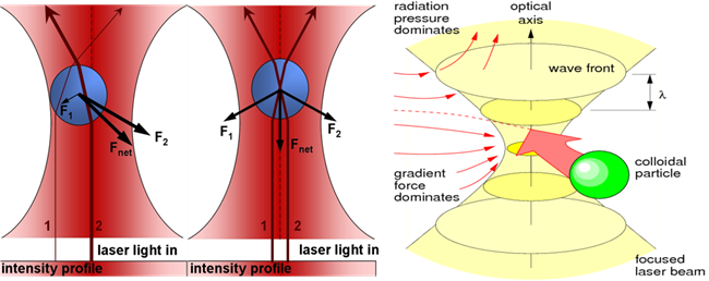
OBJECTIVE: When a light having a transverse
Gaussian mode is focused, it generates a harmonic potential field that creates
force of the ~pN range that can trap ~ micron sized particles. It has a wide
range of applicability starting from physical sciences, biological sciences,
chemical sciences etc.
THEORY: On tight focusing the
harmonic potential, generated by the Gaussian laser beam, attracts the
particles towards its focus. For
this tweezing action, we need to have microscope
objective with Numerical Aperture being greater than 1.2.The ray optics diagram
of optical tweezer is given below
INSTRUMENTS REQUIRED:
1. LASER, Mirrors,
mirror holders, lenses, lens holders, irises, filter, LED light source.
2. High
numerical aperture objectives, Dichroic mirror.
3. CCD
camera, Photo Diode.
4. Laser
glasses for eye safety.
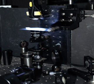
Optical Tweezer set up
SOFTWARE USED:
- LabVIEW software for data
acquisition.
- Origin pro for data plotting and analysis.
Note: For user
operation and usage no specific software needed.
EXPERIMENT PROCEDURE:
1. Make
a through hole on a microscopic slide.
2. On
one side of the hole stick a no.1 cover slip (0.13 � 0.17 μm thickness) such
that it does not come out easily from the slide. Thus a well is being formed.
3. Get
~ 1 μM concentration of ~1μm diameter polystyrene microsphere coated
with fluorophores dispersed in water.
4. Sonicate
the sample in (3) for ~ 20 minutes on a bath sonicator.
5. Turn
on the trapping Laser.
6. Turn
on the �ML� button to convert it to the femtosecond pulse Laser.
7. Place
one drop this sonicated said sample in (3) on the well.
8. Place
a drop of immersion oil on the oil immersion microscope objective with
Numerical Aperture being greater than 1.2.
9. Place
the microscopic slide with the sample being placed on the well made onto it on
the sample plane.
10. Tighten
it with the clamps.
11. Move
the objective such that the oil placed on it touches the bottom side of the
slide.
12. Let
the femtosecond Laser pulses pass through the objective and get focused in the
bulk sample (i.e., microscopic slide with the sample being placed on the well
made onto it).
13. Fix
the power of the Pulsed Laser beam.
14. Turn
on the CCD camera to record the trapping event.
15. When
the ~1μm diameter polystyrene microsphere coated with fluorophores comes
in vicinity of the focus, it feels the optical force field and jumps into the
focus and as the microspheres are coated with flourophores, so the trapped
microspheres generates the two-photon fluorescence denoting a microsphere is
being trapped.
16. CCD
camera records the trapping event that is described in process (11).