Experimental Facilities in our laboratory
- Zygo Optical Profilometer
- Optical Microscope
- Atomic Force Microscope
- High Frame Rate Camera
- Syringe Pumps
- Spin coater
- Vibration Isolation Table
- Motorized motion control stages
- Load cell, Amplifier and Data Acquisition System
- High Vacuum Unit, Plasma Oxidizer/cleaner, UV Ozonizer
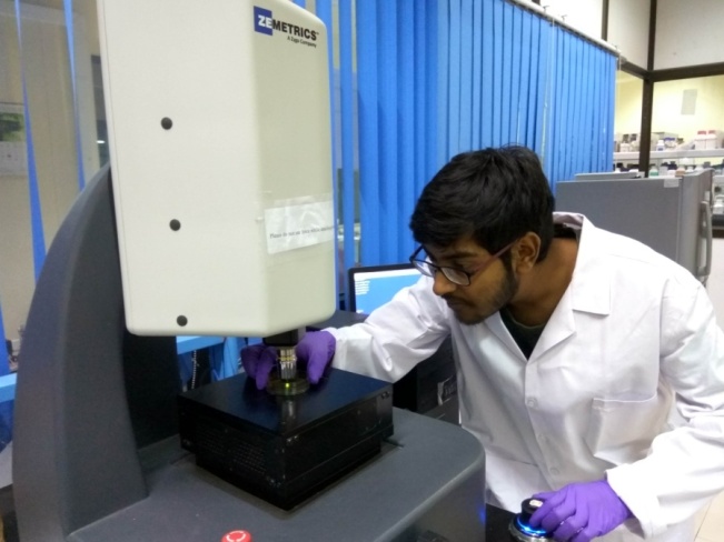
Zygo Optical Profilometer
We have in our lab a state of the art optical profilometer which helps us capture the 3-dimensional topography of microscopic features on a surface in a non-contact method. It is a work horse in our laboratory which is used by almost all students of our lab. We help also students from other laboratories in the institute and even from some start-up companies to carry out scanning of their surface.
Optical Microscope:
We have in our lab theNIKON Inverted Research Microscope Model TE2000-U that allows viewing under white light and also fluorescent light. We have 4x, 10x, 20x, 40x and 100x lens and using these lenses we can magnify a microscopic object by 150 times.
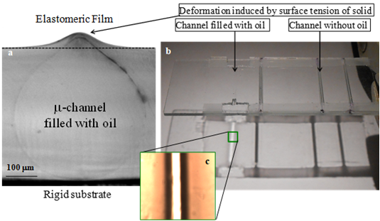
Microchannels embedded inside a thin elastomeric film are filled with an oil that wets its surface. Wetting induces surface deformation of the embedded channel. Optical microscopy captures the cross-section of the channel and also top view [Roy et al, Adv. Opt. Mat. 2014; Langmuir 2016; APL 2017].
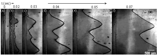
Evolution of adhesion induced instability in a thin, uniaxially stretched elastic film. The experiment corresponds to Ghatak et al 2000 (PRL). Initially there remain waves of all different wavelengths, which evolve to form well defined V
shaped fingers (Unpublished results).
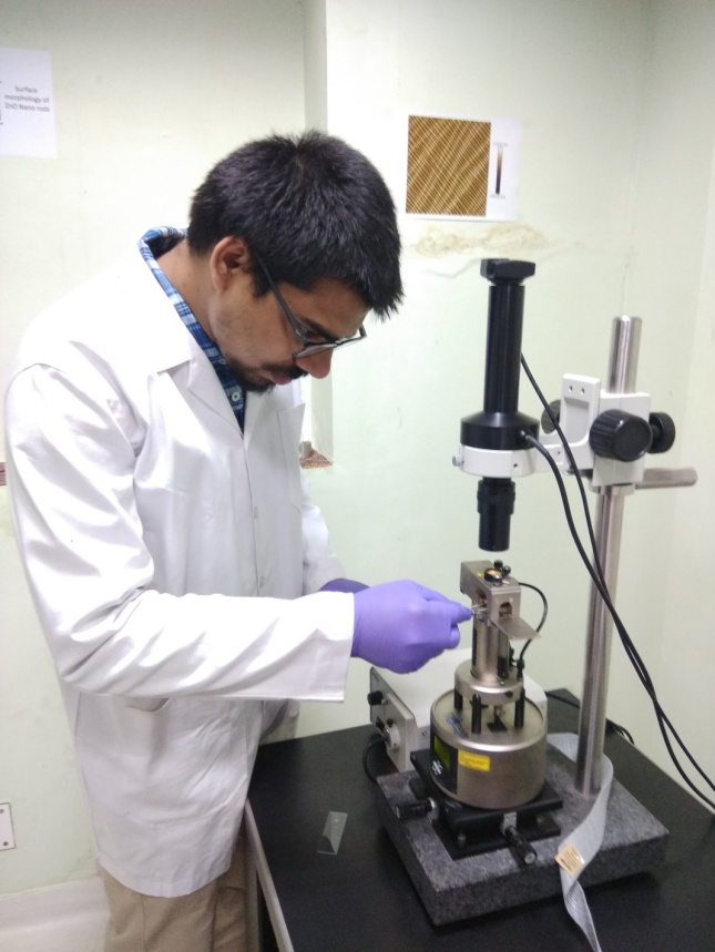
Atomic Force Microscope: Bruker Multimode 8
We have in our lab a state of the art Atomic Force Microscope which we use for variety of purposes, e.g. scanning of roughness of surface, measurement of surface potential and measurement of distribution of modulus of a soft sample.
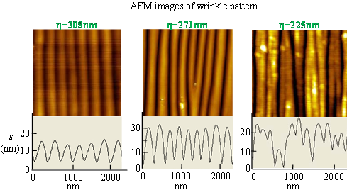
PDMS films are stretched, surface plasma oxidized and is then relaxed instantaneously. A thin crust of silica forms because of plasma oxidation which buckles when the film is relaxed, leading to the appearance of wrinkles [Sengupta et al, J. Cryst. Grow. Des., 2016; Langmuir 2013,I&EChR2011].
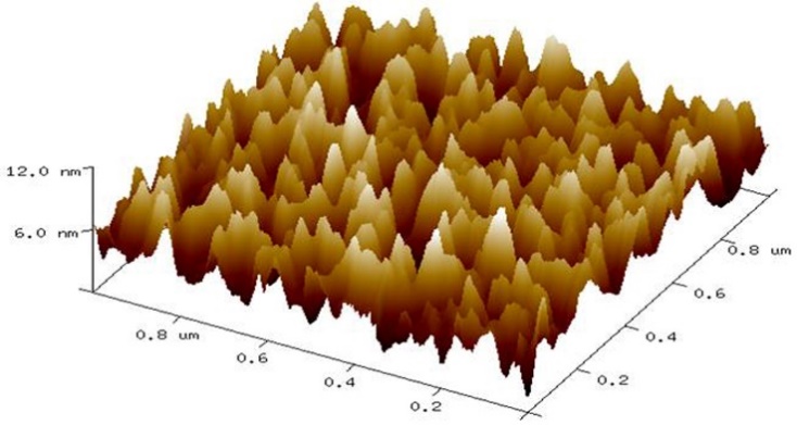
AFM scan of nanoscopic roughness of a soft surface (unpublished results).
High Frame Rate Camera
We have two high frame rate cameras, one for black and white and one for color imaging. Both these cameras allow capturing a video at 2000 fps at maximum resolution and at 100000 fps when the resolution is minimum. These cameras can be linked to our optical microscope for capturing microscopic phenomenon. The cameras are interfaced with also multiple computers.
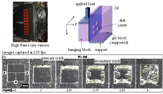
A gel block consisting of two separate parts joined via one or an array of gel disks, all prepared monolithically is subjected to shear load. At this the gel block undergoes fracture (nearly) along plane of the disk. The high frame rate camera is used to capture the fracture phenomenon [Kundan et al, 2020, PRE].
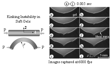
A gel rod is axially compressed leading to its buckling. The buckled rod is subjected to further bending leading to the appearance of a kink at the compressed side of the cylinder[Ghatak et al, 2006, PRL]
Syringe Pumps
We have syringe pumps for carrying out microfluidic experiments. These pumps are amenable for interfacing with computer and also with PIV equipment.
Spin coater:
We have several types of equipment for making thin films, e.g. Spin coater and Screen Printer. The former is used for experimental purposes. The later machine is useful for our scale up work.
Motorized linear stages, vibration isolation table, load cell, amplifier and data acquisition System
For carrying out load vs. displacement experiments related to adhesion and fracture tests that we do regularly, our lab is equipped with vibration isolation tables, load cell, amplifier and data acquisition system.
High Vacuum Unit, Plasma Oxidizer/cleaner, UV Ozonizer
These equipmentare used for our experiments related to surface modification of glass and siliconwafer.