FLUORESCENCE MICROSCOPE
NPTEL: https://www.youtube.com/watch?v=yZKdFVAJcrE&list=PLyAZSyX8Qy5AOfbyCYeI6n9mpMW4QLnO2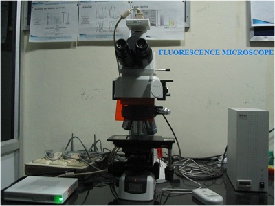
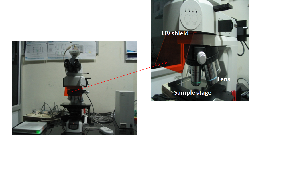
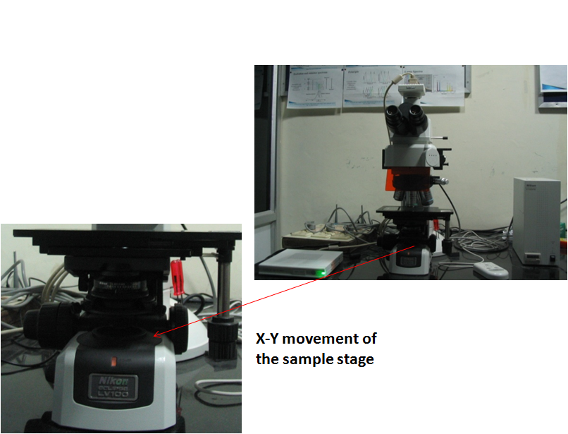
To view the various parts of the microscope click the following:
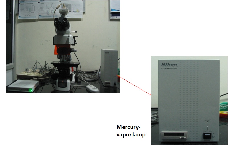
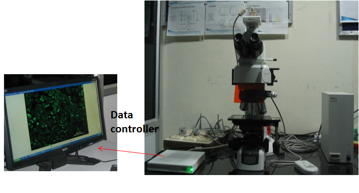
Digital Camera & Eye piece UV Shield,Lens,Sample Stage X-Y Moment of the Sample Stage Mercury Vapor Lamp Data Controller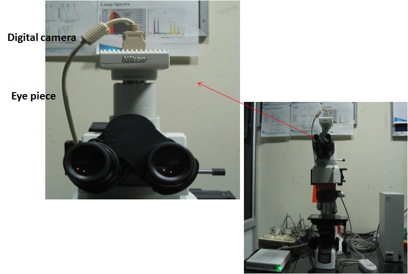
Principle
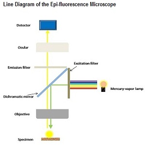
The emission of light of higher wavelength after the irradiation of the sample with known specific wavelength within nanoseconds is called Fluorescence.An instrument called fluorescence microscope is an optical microscope which utilizes a specific wavelength of light to study the properties of substances by the principle of fluorescence. Monochromatic light is used to illuminate the specimen which excites the fluorophore molecules. These fluorophore molecules release low intensity light of higher wavelengths during relaxation which passes through dichromatic mirror and emission filter which blocks the excitation light and allows the emitted light to pass, and enhances the intensity of the emitted light. Hence, single colure image can be obtained at a time due to the excitation of single fluorophore. A sample containing different fluorophore are imaged to observed different features present in the sample and these single colored images are merged to obtain the multi-colored images.
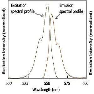
Excitation and Emission Spectra
(1) The excitation and emission spectra are nearly the mirror image of each other.
(2) Emission spectra shifts towards higher wavelengths.
(3) The difference between the wavelength is called Stoke's Shift.
(4) The intensity of emitted light is generally of 10-6 times of excitation light.
Precautions
(1) Turn-off the ligths of the room during the fluorescence experiment.
(2) Do not expose the fluorophore treated specimen under the specific wavelength of light as the duration of the fluorescence phenomena is of the order of 10-6-10-9 seconds.
(3) Always use the UV shield while using the eyepiece.
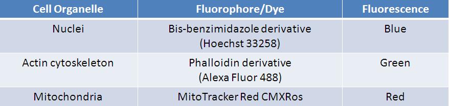
Common Staining Agents used in Biological Studies
Applications
Biological and Life Sciences
(1) To view tissue and their sub-microscopic components and changes their in.
(2) To study bacteria and pathogens and changes their-in.
(3) Protein-protein and protein-cell interaction study.
(4) Movement of virus on a bi-layered membrane.
(5) Crude drugs.
Material Science
To see the various attributes of minerals, crystals, resins, chlorophyll and inorganic compounds.